Every time you pick up a cup, climb a staircase, or take a breath, millions of microscopic protein filaments inside your muscle fibers are sliding past each other in a precisely coordinated sequence. That process is described by the sliding filament theory, and it’s one of the most elegant explanations in all of biology. It takes something as familiar as a muscle contraction and reveals the astonishing molecular machinery running underneath.
The sliding filament theory was proposed in 1954 by two independent research teams — Andrew Huxley and Rolf Niedergerke, and Hugh Huxley and Jean Hanson — working separately but arriving at essentially the same conclusion at the same time. Their insight was that muscle fibers don’t shorten because the proteins inside them get smaller. The proteins stay the same length. Instead, they slide past each other, overlapping more during contraction than during rest. That single idea reorganized how scientists thought about muscle physiology and laid the groundwork for everything that followed in the field.
Muscle Fiber Basic Structure
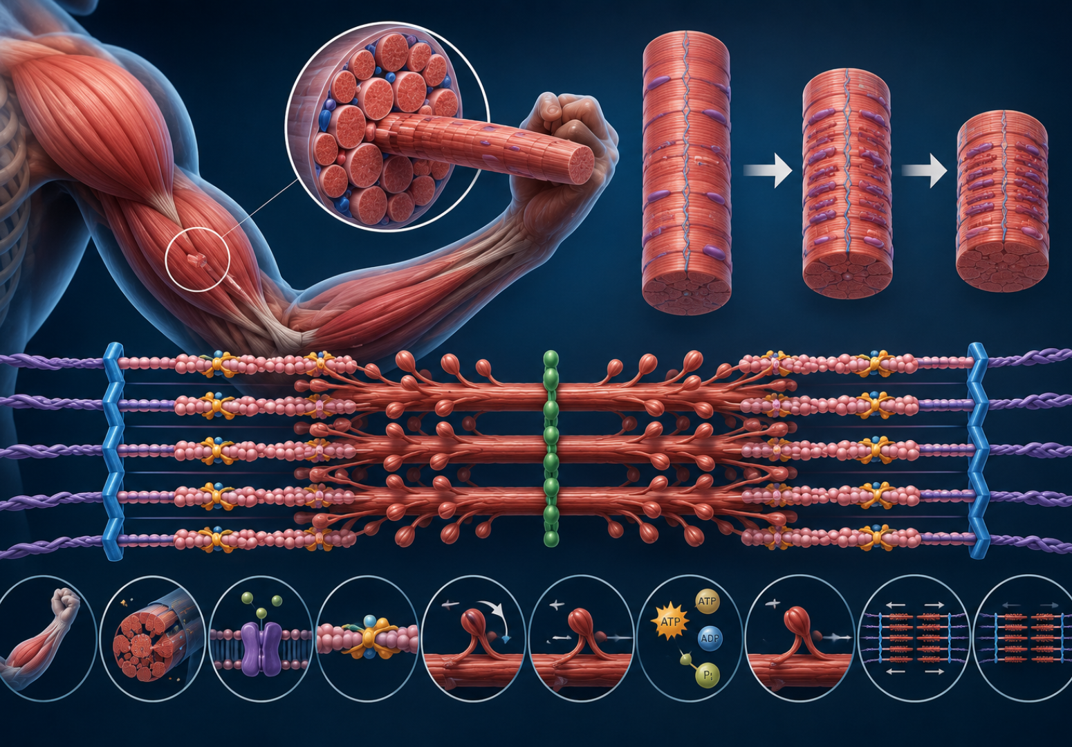
Before the sliding filament theory makes full sense, you need a clear picture of what a muscle fiber actually looks like at the microscopic level. A single skeletal muscle is made up of bundles of muscle fibers, and each fiber is a long cylindrical cell containing hundreds of even smaller structures called myofibrils. These myofibrils run the entire length of the fiber and are the actual contractile units of the muscle.
Each myofibril is divided into repeating segments called sarcomeres, which are the fundamental units of muscle contraction according to the sliding filament theory. A sarcomere runs from one Z-line to the next, spanning roughly 2.2 micrometers at rest. Inside each sarcomere are two types of protein filaments arranged in a specific geometric pattern — thick filaments made of myosin and thin filaments made primarily of actin. The way these filaments are arranged creates the characteristic banded appearance of skeletal muscle under a microscope, which is why it’s called striated muscle. When scientists first observed these bands in the 19th century, they had no idea they were looking at the physical basis of movement. The sliding filament theory is what finally explained the pattern.
Actin and Myosin Proteins
The two main players in the sliding filament theory are actin and myosin, and they couldn’t be more different in structure and function. Actin forms the thin filaments. It’s a globular protein (G-actin) that polymerizes into long double-stranded helical chains (F-actin), giving the thin filaments their characteristic twisted-rope appearance. Wound around the actin helix are two regulatory proteins — tropomyosin, which runs along the groove of the helix, and troponin, a complex of three subunits that controls whether contraction can happen.
Myosin forms the thick filaments and is the molecular motor of the sliding filament theory. Each myosin molecule has a long tail region and two globular head domains that project outward from the thick filament. These heads are the business end of the whole operation — they bind to actin, hydrolyze ATP, and generate the force that drives filament sliding. A single thick filament contains around 300 myosin molecules, all arranged so their heads point outward and away from the center of the sarcomere. When conditions are right, those heads grab the actin filaments on either side and pull them inward. That’s the contraction. That’s the sliding filament theory in its most distilled form. For teachers who want to bring this kind of cellular biology to life in classrooms, science education engagement offers some genuinely useful perspectives on making complex science accessible.
The Cross Bridge Cycle
The cross bridge cycle is the molecular sequence that actually drives contraction in the sliding filament theory, and it’s worth walking through step by step because each stage is mechanistically distinct. It begins when a myosin head, already energized by ATP hydrolysis, binds to an exposed actin binding site on the thin filament. This binding is the “cross bridge” — the physical connection between thick and thin filaments.
Once bound, the myosin head undergoes a conformational change called the power stroke. It pivots at its hinge region, pulling the actin filament toward the center of the sarcomere by about 5 to 10 nanometers. This movement is tiny but it happens simultaneously across hundreds of sarcomeres in series and thousands of myosin heads in parallel, so the cumulative effect is a visible, forceful muscle contraction. After the power stroke, a new ATP molecule binds to the myosin head, causing it to release from actin. The ATP is then hydrolyzed back to ADP and inorganic phosphate, re-energizing the head and resetting it for another cycle. This cycle repeats as long as calcium ions are present and ATP is available — the two essential conditions for sustained contraction under the sliding filament theory.
Role of Calcium Ions
Calcium is the switch that turns muscle contraction on and off, and its role is central to the sliding filament theory. At rest, tropomyosin physically blocks the myosin binding sites on actin filaments. The cross bridge cycle can’t start because myosin heads have nowhere to attach. Then a nerve signal arrives.
When a motor neuron fires an action potential that reaches the neuromuscular junction, it triggers the release of acetylcholine, which depolarizes the muscle fiber membrane. That depolarization travels into the fiber along structures called T-tubules and reaches the sarcoplasmic reticulum — a specialized network of membrane-enclosed compartments wrapped around each myofibril. The sarcoplasmic reticulum responds by releasing stored calcium ions into the cytoplasm surrounding the myofibrils. Calcium binds to troponin C, the calcium-sensing subunit of the troponin complex. This binding causes a conformational change in troponin that shifts tropomyosin away from the myosin binding sites on actin — exposing them. The cross bridge cycle can now begin. The sliding filament theory would be incomplete without this calcium-mediated regulatory mechanism, because it explains not just how muscles contract but how contraction is controlled with such precision.
ATP as the Energy Source
ATP is the fuel that powers every step of the sliding filament theory, and muscle cells have several systems for producing it quickly enough to sustain contraction. During the cross bridge cycle, each myosin head uses one ATP molecule per cycle. Given that a single muscle fiber contains millions of myosin heads cycling many times per second during intense activity, the ATP demand is enormous.
Muscle cells meet this demand through three overlapping energy systems. The phosphocreatine system provides ATP almost instantly by transferring a phosphate group from creatine phosphate to ADP, but it depletes within 10 seconds of maximum effort. Anaerobic glycolysis then kicks in, breaking down glucose to pyruvate (and then lactate) to produce ATP without oxygen — fast but inefficient, producing only 2 ATP per glucose. For sustained activity, aerobic respiration in the mitochondria takes over, producing up to 36-38 ATP per glucose molecule. The accumulation of metabolic byproducts during intense exercise — including inorganic phosphate, hydrogen ions, and ADP — is thought to contribute to muscle fatigue by interfering with the cross bridge cycle described in the sliding filament theory. The exact mechanisms of fatigue are still an active research area.
History of the Theory
The story of how the sliding filament theory came to be is worth knowing, not just as trivia but because it illustrates how science actually works. By the early 1950s, it was known that muscles contained actin and myosin and that these proteins were somehow involved in contraction. The prevailing idea was that the proteins themselves shortened — folded or coiled up — during contraction. This seemed logical but turned out to be wrong.
In 1954, two papers published back-to-back in the same issue of Nature changed everything. Andrew Huxley and Rolf Niedergerke used interference microscopy to observe living muscle fibers contracting and relaxing. Hugh Huxley and Jean Hanson used electron microscopy to examine the fine structure of myofibrils. Both pairs independently observed that the A-band (the region containing thick filaments) stayed constant in width during contraction, while the I-band (the region containing only thin filaments) narrowed. If the proteins themselves were shortening, you’d expect the A-band to shrink too. It didn’t. The only explanation that fit was filament sliding — and the sliding filament theory was born from two independent observations converging on the same answer simultaneously.
Neuromuscular Junction Explained
The neuromuscular junction is where the nervous system hands off control to the muscle, and it’s the starting point for every contraction described by the sliding filament theory. According to research published by the National Institutes of Health, the neuromuscular junction is one of the most studied synapses in the body, partly because of its accessibility and partly because its dysfunction underlies diseases like myasthenia gravis and Lambert-Eaton syndrome.
At the neuromuscular junction, the terminal of a motor neuron sits close to a specialized region of the muscle fiber membrane called the motor end plate. When an action potential reaches the terminal, voltage-gated calcium channels open, calcium flows in, and synaptic vesicles fuse with the membrane, releasing acetylcholine into the synaptic cleft. Acetylcholine binds to nicotinic receptors on the motor end plate, generating an end plate potential that — if large enough — triggers an action potential in the muscle fiber. That action potential is what initiates the calcium release from the sarcoplasmic reticulum and sets the sliding filament theory in motion. The whole sequence from nerve signal to filament sliding takes just a few milliseconds.
Sliding Filament Theory in Smooth Muscle
Most discussions of the sliding filament theory focus on skeletal muscle, but smooth muscle — the kind lining your blood vessels, digestive tract, airways, and uterus — uses the same basic mechanism with some important differences. Smooth muscle cells don’t have the organized sarcomere structure of skeletal muscle. There are no visible striations, no Z-lines, no neat arrangement of thick and thin filaments into regular repeating units.
Despite this, the sliding filament theory still applies. Smooth muscle contains actin and myosin that slide past each other during contraction. The regulation is different — smooth muscle uses calmodulin rather than troponin as its calcium sensor, and myosin must be phosphorylated by an enzyme called myosin light chain kinase before it can bind actin. Smooth muscle also contracts more slowly than skeletal muscle and can sustain contraction for much longer without fatigue, which suits its role in maintaining prolonged states like blood vessel tone and digestive peristalsis. The sliding filament theory is flexible enough as a framework to accommodate these differences while still describing the core mechanism accurately.
Cardiac Muscle Contraction Differences
Cardiac muscle is its own category — striated like skeletal muscle but involuntary like smooth muscle — and the sliding filament theory applies here too, with a few cardiac-specific twists. The most important is that cardiac muscle cells are electrically coupled through structures called gap junctions, so the heart contracts as a coordinated unit rather than requiring individual motor neuron input for each cell.
Cardiac muscle also has a longer action potential than skeletal muscle — about 200 to 300 milliseconds compared to 1 to 2 milliseconds — which produces a prolonged calcium release and a longer contraction. This long contraction period is essential for pumping blood effectively. The heart can’t tetanize (remain in sustained contraction) the way skeletal muscle can, and that’s protective — a heart in sustained contraction wouldn’t pump blood at all. Within cardiac muscle cells, the cross bridge cycle described by the sliding filament theory operates the same way as in skeletal muscle, but the regulatory mechanisms that control when and how strongly it operates are tuned very differently to meet the heart’s unique demands.
Muscle Fatigue and the Theory
Muscle fatigue — the decline in force production during sustained or repeated contraction — is one of the most practically relevant aspects of the sliding filament theory, and it’s more complex than most people realize. The popular notion that lactic acid causes fatigue has been largely revised by modern research. Lactate itself isn’t the villain; it’s the accompanying hydrogen ions from ATP hydrolysis and glycolysis that lower intracellular pH and interfere with the cross bridge cycle.
At the level of the sliding filament theory, fatigue manifests as a reduction in the number of cross bridges that can form and a slowing of the cross bridge cycling rate. Accumulation of inorganic phosphate (released when ATP is hydrolyzed to ADP) interferes with the power stroke step. Reduced calcium release from the sarcoplasmic reticulum means fewer binding sites are exposed on actin. The myosin heads themselves may become less responsive to calcium under fatiguing conditions. All of these effects reduce the force generated per sarcomere, and when that happens across millions of sarcomeres in a working muscle, you feel it as fatigue — the burn, the weakness, the inability to maintain the same effort level. The sliding filament theory gives you the language to understand exactly where in the process things are breaking down.
Length-Tension Relationship
The length-tension relationship is one of the most important practical predictions of the sliding filament theory, and it has direct implications for how muscles work most effectively at different joint positions. The basic principle is that the force a sarcomere can generate depends on how much overlap exists between its thick and thin filaments.
At optimal length — roughly 2.2 micrometers per sarcomere in human skeletal muscle — the overlap between actin and myosin allows the maximum number of cross bridges to form simultaneously, producing maximum force. Stretch the sarcomere beyond this point and the filaments pull apart, reducing overlap and therefore reducing the number of cross bridges that can form. Force drops. Shorten the sarcomere beyond optimal length and the thin filaments from opposite sides of the sarcomere start to overlap each other and bump into the thick filaments, creating mechanical interference that also reduces force. There’s a sweet spot, and the sliding filament theory predicts it precisely. Sports scientists and physical therapists use the length-tension relationship when designing rehabilitation programs and training protocols, because joint angle determines muscle length, which determines where on the curve a muscle is operating.
Sliding Filament Theory in Disease
Diseases that disrupt any step of the sliding filament theory tend to produce significant muscle dysfunction. Muscular dystrophies, for example, result from mutations in structural proteins that support the sarcomere during the mechanical stresses of repeated contraction. Dystrophin, the protein absent in Duchenne muscular dystrophy, links the actin cytoskeleton to the extracellular matrix, providing mechanical stability. Without it, the repeated force of cross bridge cycling tears the muscle membrane, leading to progressive muscle damage.
Myosin mutations cause hypertrophic cardiomyopathy, the most common inherited heart condition, affecting roughly 1 in 500 people. Specific mutations in the beta-myosin heavy chain alter cross bridge kinetics — typically making myosin bind more tightly or cycle more slowly — which changes the mechanical properties of cardiac muscle in ways that ultimately remodel the heart’s structure. Troponin mutations can cause both hypertrophic and dilated cardiomyopathy. Understanding exactly how these mutations perturb the cross bridge cycle as described by the sliding filament theory has been essential for developing targeted therapies, including the drug mavacamten, which directly modulates myosin ATPase activity and is the first drug to target the molecular machinery of the sliding filament theory specifically.
Modern Research Developments
The sliding filament theory as originally proposed in 1954 has held up remarkably well, but modern research has added significant depth and nuance to the original model. Single-molecule techniques developed since the 1990s — particularly optical tweezers experiments — have allowed researchers to measure the force and displacement produced by individual myosin molecules during a single power stroke. The results confirmed the basic sliding model while revealing details about cross bridge mechanics that couldn’t have been measured with the tools available to the Huxleys.
Structural biology has contributed high-resolution structures of actin, myosin, and the troponin-tropomyosin regulatory complex in multiple states, giving researchers a near-atomic-level picture of what happens during each step of the cross bridge cycle. Cryo-electron microscopy has been particularly powerful here, capturing myosin heads in their pre-power-stroke and post-power-stroke conformations and revealing how ATP binding and hydrolysis drive the mechanical changes in the head domain. These advances haven’t overturned the sliding filament theory — they’ve confirmed and extended it, adding molecular detail to a framework that was already correct in its essential features.
FAQ
What is the sliding filament theory in simple terms?
The sliding filament theory explains how muscles contract by describing the movement of two types of protein filaments — actin (thin) and myosin (thick) — past each other inside muscle fibers. The filaments themselves don’t get shorter; they slide over one another, increasing overlap and shortening the sarcomere. This sliding is driven by myosin heads grabbing actin, pulling it inward, releasing, and repeating in a cycle powered by ATP.
Who proposed the sliding filament theory and when?
The sliding filament theory was proposed in 1954 by two independent research groups. Andrew Huxley and Rolf Niedergerke published one paper, while Hugh Huxley and Jean Hanson published another — both in the same issue of Nature. Both groups reached the same conclusion using different microscopy techniques, and their simultaneous discovery gave the theory immediate credibility in the scientific community.
What role does calcium play in the sliding filament theory?
Calcium is the regulatory switch. At rest, tropomyosin blocks myosin binding sites on actin, preventing cross bridge formation. When a nerve signal triggers calcium release from the sarcoplasmic reticulum, calcium binds to troponin and shifts tropomyosin away from the binding sites, allowing myosin heads to attach to actin and begin the cross bridge cycle. When the nerve signal stops, calcium is pumped back into the sarcoplasmic reticulum and contraction ceases.
How does ATP relate to the sliding filament theory?
ATP is essential at two points in the cross bridge cycle. First, ATP hydrolysis (to ADP and inorganic phosphate) energizes the myosin head and cocks it into the high-energy position ready to bind actin and perform the power stroke. Second, binding of a fresh ATP molecule to myosin after the power stroke causes the head to release from actin, allowing the cycle to repeat. Without ATP, myosin stays locked to actin — which is exactly what happens in rigor mortis after death, when ATP production stops entirely.
Conclusion
The sliding filament theory is one of those scientific ideas that genuinely changes how you see the world once you know it. Every movement your body makes — from the blink of an eye to the sustained effort of running a marathon — traces back to myosin heads grabbing actin filaments and pulling them inward, a few nanometers at a time, billions of times per second across millions of muscle fibers working in coordination. The sliding filament theory puts a precise molecular mechanism behind what used to be just an observable fact about biology.
What makes the theory particularly satisfying is how well it has survived contact with better technology. When the Huxleys proposed it in 1954, they were working with light microscopes and early electron microscopes. Seventy years later, with single-molecule force measurements and near-atomic structural data, the core model is still standing — refined and deepened, but not overturned. The sliding filament theory remains the organizing framework for muscle physiology research, for understanding muscle diseases, and for developing treatments that target the molecular machinery of contraction directly. That durability is the mark of a genuinely good scientific theory, and this one has earned its place at the center of how we think about muscle biology.